Full Text:
This full article is available in French. The following is a Google translation:
Summary
The oral cavity is one of the major routes of environmental contamination known to be involved in many chronic pathologies (cancers, fertility and behavioral disorders) viafood, medication or even breathing. These environmental factors including, inter alia, endocrine disruptors and excess fluorine, can disturb dental development and thus generate irreversible defects in the enamel. These defects are then treated with materials, some of which release molecules capable in turn of generating these defects, leading to a vicious circle, especially in pregnant women and young children. This synthesis takes stock of the state of knowledge, questions and controversies on common environmental factors likely to come into contact with the oral sphere, their mechanisms of action and the mediators involved in enamel pathologies associated with environmental conditions.
Abstract
The oral cavity is one of the main route for environmental contaminations associated to many chronic diseases (cancers, fertility and behavior disorders for example) viadiet, medications and breathing. These environmental factors including, among others, endocrine disruptors and excessive fluoride can disrupt dental development and thus generate irreversible enamel defects. These defects are then treated with materials that may release molecules capable of generating these defects, leading to a vicious circle, particularly in pregnant women and young children. The present paper aims to review the state of knowledge, questions and controversies on common environmental factors in contact with the oral cavity. It also reviews their mechanisms of action and the mediators involved in enamel pathologies associated with environmental conditions. Dental tissues can not only be targeted by environmental factors but can also serve as early and easily accessible markers of exposure to these agents.
© 2020 medicine / science – Inserm
Article published under the conditions defined by the Creative Commons Attribution License CC-BY license ( https://creativecommons.org/
Dental tissue can not only be the target of environmental factors but also serve as early markers and is easily accessible and exposed to these agents. Understanding and characterizing the environmental impact in the oral sphere will help prevent multiple pathologies not only in this cavity, but also more distant pathologies whose link with oral homeostasis is just beginning to be explored.
Contaminating environmental factors
The oral cavity is contaminated by a variety of environmental factors which, for some, are grouped under the term of “food contaminants”, when individuals do not identify them and undergo their contamination: for example, the mycotoxins produced by fungi. Others, called “toxic”, correspond to contaminants that have been knowingly consumed, knowing their side effects or the risk associated with their consumption, such as alcohol, tobacco, certain drugs, even sugar when taken in excess. Contamination of a food can also occur during the various stages of its production: the foodstuffs produced may in fact have been originally contaminated by the presence of compounds involuntarily introduced into the soil (for crops) or into the food chain (for livestock and industrial production), such as dioxins, biocides, plasticizers , among others. Phytosanitary products (or pesticides including fungicides, herbicides, insecticides, etc.) are themselves commonly used during the production process: harvesting, conservation of fruits, vegetables and cereals (which will be used or not for the breeding). The processes used to make the products (smoking and cooking) can also generate neo-synthesis of molecules which prove harmful to health, such as polycyclic hydrocarbons (PAH) or acrylamide, now identified as a carcinogen. Acrylamide is produced by the glycation reaction of products when they are heated or cooked. The packaging processes, on the other hand, can cause the transfer of bisphenols and phthalates from the packaging product to the food. All of these xenobiotics are strongly suspected of inducing metabolic, malignant and autoimmune pathologies in adults, but also developmental pathologies affecting the fetus,via the mother, or the young child. The prevalence of these non-infectious pathologies being in constant increase since the last decades, in particular in the young subjects, the knowledge, even the control, of these etiological factors thus appear essential. The questions which relate to their capacity for disturbance and their mechanisms of action within the oral cavity thus legitimately arise and should be the subject of specific research in the coming years.
Dental enamel marker of environmental conditions
Dental development and, in particular, the synthesis of enamel follow a sequence of molecular and cellular events perfectly ordered in space and time [1 ]. There are three major phases in amelogenesis 1 : the proliferation and recruitment of stem cells; secretion, during which the enamel matrix proteins determine the thickness of the enamel and help in its mineralization; and maturation which sees almost all of these amelar proteins degraded to leave the space made free for the growth of apatite crystals and the complete mineralization of enamel [2 ] ( ? ), the most mineralized tissue in the body covering the dental crown. When this process ends, the tooth is ready to erupt in the oral cavity. The ameloblasts are lost during this last stage, which makes any future damage to the enamel irreparable and therefore irreversible. Thus, exogenous disturbances affecting ameloblasts or endogenous regulations of their activity and survival, can lead to amelar stigmas, witnesses of these disturbances. These stigmas can thus help to reconstruct the history of exposures and contaminations that the ameloblasts have undergone ( Figure 1 ) .
– See the Synthesis by G. Lignon et al ., m/s n° 5, May 2015, page 515
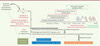 |
Figure 1.Exposure window and selectivity of damage to dental enamel. Exposure to various environmental factors can alter the activity of ameloblasts, cells responsible for the synthesis of enamel, and generate irreversible enamel defects on the teeth that form during this period of exposure. Dental development extends from fetal life to 4-7 years after birth, a period of maximum susceptibility to environmental toxins, the exposure of which can be decisive for the state of health in adults. The resulting amelar hypomineralizations are treated with materials which can release bisphenol monomers which in turn may contribute to the development of disease. The tooth can therefore serve as an early marker of exposure to these agents. |
The development of each tooth has been described in detail over time [3 ]. This allows the window of exposure time to agents which have selectively impaired the development of certain teeth to be defined fairly precisely. The development of dental enamel takes place during the last two trimesters of fetal life and between 4 and 6 years after birth, which is a decisive window of time for the adult’s future state of health [4 ]. All of these unique and unique characteristics of enamel give this tissue properties of tracer of exposure to agents which could disturb its synthesis, but also alter the health of an individual, as seems to be the case with disruptors. endocrine [5 ].
How can certain endocrine disruptors cause enamel defects?
The concept of “endocrine disruptor” emerged in the early 1990s following the Winspread 2 conference [6]. Re
Massive contamination of populations (over 90% of individuals) has been demonstrated for the dozens of the most widespread (or most sought after) PEs [11 ]. And many recent epidemiological studies establish a link between the degree of contamination of populations and the incidence of genital tract malformations, obesity, behavioral disorders, certain hormone-dependent cancers (to mention only the studies more common) [ 5]. Exposure to PE is the main hypothesis explaining the worrying decline in male fertility worldwide. Their activity is no longer discussed today in the scientific and medical community; it has been widely demonstrated experimentally. The current challenges aim to establish precisely the links between the results obtained experimentally on animals and epidemiological data, and relate to the health impact of combinations of PE when used in low doses (cocktail effect).
Among the 1,491 molecules with PE activity (referenced in the TEDX List of June 2019), bisphenol A (BPA) appears to be exemplary, as it has been studied under various experimental and clinical conditions. It is also one of the only non-medicinal manufactured molecules classified by the European Food Safety Authority (EFSA) as a proven PE for health (in 2017) and the environment (in 2018). Numerous experimental data obtained in rodents make it possible to identify the pathological consequences linked to exposure to BPA, consequences which are probably identical in humans. So in rats,12 ], a pathology of enamel described only in 2001 [13 ] and whose etiological factors are still unclear [14 ]. In this pathology, the affected teeth present localized opacities reflecting a hypomineralization whose severity can be assessed: the teeth developing first are preferentially affected, the enamel is porous and capable of accumulating albumin ( Figure 1 ) . PEs whose activity is similar to that of BPA therefore prove to be interesting candidates as factors potentially capable of generating MIH. Studies support this hypothesis, in particular those showing hypomineralizations of the enamel following the chronic exposure of rodents to vinclozoline, genistein, phthalates or dioxin ( Figure 1 ) [15 ,16 ], or associating hypomineralizations of the enamel and contamination by dioxin and by polychlorinated biphenyls (PCB) in humans [ 16 ,17 ]. The comparison of dental defects obtained after exposure to these different PE reveals specific pathophysiological characteristics, suggesting a mode of action and cellular and molecular targets specific to each PE or to each mixture of PE. These differences thus raise questions about the heterogeneity of MIH: this pathology could, in fact, group together different enamel anomalies, affecting a wider variety of teeth than that initially proposed [18 ]. This heterogeneity could originate from variable combinations of exposure to causative agents, grouping not only PEs but also, possibly, antibiotics and drugs in a particular environmental and genetic context, still unknown, and which would favor the action of these molecules [ 14 ,19 ].
Ameloblasts express most of the steroid hormone receptors [20 ], those who are involved in the pathophysiological effects of PE. Moreover, the synthesis of enamel is modulated by androgens [21 ] and with vitamin D [22 ]. Retinoids, metabolites of vitamin A, have also been implicated in the regulation of amelogenesis [23 ]. It has in fact been shown that mice exposed during fetal life to an excess of retinoic acid have a reduced quantity of dental enamel and a very altered bone.
Understanding the mechanisms of action of these disruptive molecules should make it possible to put an end to the controversy between researchers, industrialists, politicians and health agencies because of contradictory economic and ideological issues [24 ]. Pending an objective analysis, the health costs linked to the diseases associated with exposure to these PEs have been evaluated [25 ]: the only prenatal exposure to BPA was identified as probably associated with 42,400 cases of childhood obesity in Europe, with an overall cost estimated at 1.54 billion euros.
The oral cavity sits in a vicious circle
Certain dental materials commonly used in conservative dentistry and for orthodontic treatments release BPA monomers ( Figure 1 ) [26 ]. An awareness of dentists asking manufacturers for a list of the constituents of the materials they used, led them to seek new formulations and procedures for use tending to reduce the amount of BPA monomers potentially released. None of these biomaterials contain pure BPA, but most of them are synthesized from monomers which are derived from BPA, or from other monomers which may have cytotoxicity [27 ]. It is therefore essential to know the composition and to master the protocols for the use of these materials which are commonly used for the treatment of caries, in particular in the case of MIH, in order to break a vicious circle which can settle down between exposure of the patient. and treatment of its diseases which are themselves associated with this exposure, especially since these molecules can exhibit non-monotonic activities, with significant effects at very low doses [28 ], and pass the sublingual barrier [29 ].
The discovery of the sublingual passage of BPA in dogs indeed questions the chronic impact of this contaminant, used in low doses, on the oral cavity. It highlights a possible systemic contamination to other target tissues, via the blood circulation [ 5]. This sublingual passage can thus occur in individuals who have been treated for cavities for several years with dental composites which were less controlled than today and handled according to procedures less well identified than at present. This possible passage into the bloodstream could explain the circulating levels of BPA which turn out to be much higher than the predicted values according to the average data of contamination and the intense metabolism of BPA into BPA glucuronide by the liver. However, the clearance of BPA remains important in humans: it is estimated that less than 0.5% of the amount ingested reaches the general blood circulation in active form. But this percentage reaches almost 60% for bisphenol S (BPS), one of the substitutes for BPA,30 ]. The substitution of BPA by BPS is therefore worrying: it risks further increasing the internal exposure of individuals. It is therefore necessary to know the mechanisms of action of these substitution molecules proposed by manufacturers in order to understand their possible effects on health.
Fluoride: beneficial effects and side effects
Fluorine is one of the molecules in chronic contact with the oral cavity. Capable of (re) mineralizing the enamel on the surface and inhibiting the bacterial enolases causing cavities, it is therefore commonly used to prevent this damage. However, exposure to excessive fluoride can lead to the development of dental and skeletal fluorosis, conditions that were identified in the first half of the XX th century [31 ]. Dental fluorosis is, like MIH, a pathology of the development of enamel, which, like the latter, causes opaque whitish to brownish spots, which can, in some cases, pose diagnostic problems between the two pathologies. Fluorosis affects less than 3% of children in France, but nearly 200 million people worldwide, distributed in 25 countries [32 ]. The dose of prophylactic fluoride is evaluated at 0.05 mg / kg / day. But a dose greater than 0.1 mg / kg / day exposes you to a risk of fluorosis. However, it is frequent that the fluorine content of drinking water, in particular certain mineral waters, is between 0.3 to 0.5 mg per liter, doses capable of inducing fluorosis.
In addition to its extracellular properties which are well documented, recent data report intracellular effects of fluorine, with the modulation of gene expression in the dental epithelium, but also in other tissues ( Figure 1 ) . Exposure to fluoride has thus been associated with effects beyond the oral framework and mineralized tissues, including neurotoxic effects and a reduction in the intelligence quotient [33 ,34 ], disturbances of the androgenic axis [35 ] (fluorine is listed as a PE in the TEDX List), and an increase in inflammatory processes [36 ]. The involvement of steroid hormone receptors (progesterone and androgens) in the mechanisms of action of fluorine on ameloblasts [37 ] thus suggests strong interactions between fluorine and PE. Moreover, fluorine and BPA can have complementary and additional effects as disruptors of amelogenesis [38 ]. This could explain the increase in sensitivity to fluoride and an increase in fluorosis in the population [39 ]. Fluoride also reduces the ability of cells to store iron, notably by reducing the quantity of heavy ferritin chains [40 ]. This action on the storage of iron can have many consequences on the cellular processes which involve it, such as oxidative stress and cell proliferation. All of this data leads to reconsider the need to supplement individuals with fluoride, taking into account changes in lifestyles and access to foods naturally rich in fluorine (tea for example).
Conclusion
Dental defects resulting from exposure to environmental agents could be used as early exposure markers, or even markers of prognosis for diseases associated with these exposures and diagnosed later, often during adulthood, while dental defects are observed as soon as possible. childhood at the time of the dental eruption. Indeed, the enamel of temporary teeth is synthesized from fetal life and that of permanent teeth, from birth to adolescence. We also know that perinatal conditions are decisive for the health of the future adult. Dental enamel could therefore be a very early reflection.
Links of interest
The authors declare that they have no link of interest concerning the data published in this article.
2The Wingspread conference in Wisconsin brought together 21 participants from July 26 to 28, 1991; it is the fruit of Theodora Colborn’s work. If the concept of endocrine disruptor was invented at Wingspread in 1991, two publications will be at the origin of its development: a report written at the request of the Danish Ministry of the Environment and Energy in 1995 and a book published in 1996 by Theodora Colborn, prefaced by Al Gore, then Vice-President of the United States, which will have a global impact. https://www.senat.fr/
References
- Lacruz RS, Habelitz S, Wright JT, Paine ML. Dental enamel formation and implications for oral health and disease. Physiol Rev 2017; 97: 939–993. [Google Scholar]
- Lignon G, from Dure-Molla M, Dessombz A, Berdal A, Babajko S. Enamel: a self-assembly unique in the mineral world. Med Sci (Paris) 2015; 31: 515-521. [CrossRef] [EDP Sciences] [PubMed] [Google Scholar]
- AlQahtani SJ, Hector MP, Liversidge HM. Brief communication: the London atlas of human tooth development and eruption. Am J Phys Anthropol 2010; 142: 481-490. [CrossRef] [PubMed] [
Google Scholar] - Bertagnolli M, Luu TM, Lewandowski AJ, et al. Preterm birth and hypertension: is there a link ?. Curr Hypertens Rep 2016; 18: 28. [CrossRef] [PubMed] [
Google Scholar] - Gore A, Chappell V, Fenton S, et al. EDC-2: The endocrine society’s second scientific statement on endocrine-disrupting chemicals. Endocr Rev 2015; 36: E1–150. [Google Scholar]
- Colborn T .. Pesticides-how research has succeeded and failed to translate science into policy: endocrinological effects on wildlife. About Health Perspect 1995; 103: 81–85. [PubMed] [Google Scholar]
- Karthikeyan BS, Ravichandran J, Mohanraj K, Vivek-Ananth RP, Samal A. A curated knowledgebase on endocrine disrupting chemicals and their biological systems-level perturbations. Science Total Environm 2019; 692: 281-296. [CrossRef] [Google Scholar]
- Corbel T, Perdu E, Gayrard V, et al. Conjugation and deconjugation reactions within the fetoplacental compartment in a sheep model: a key factor determining bisphenol A fetal exposure. Drug Metab Dispos 2015; 43: 467–476. [CrossRef] [PubMed] [
Google Scholar] - Du Z, Cao YF, Li SN, et al. Inhibition of UDP-glucuronosyltransferases (UGTs) by phthalate monoesters. Chemosphere 2018; 197: 7-13. [CrossRef] [PubMed] [
Google Scholar] - Beszterda M, Franski R. Endocrine disruptor compunds in environment: as a danger for children health. Pediatr Endocrinol Diabetes Metab 2018; 24: 88–95. [Google Scholar]
- Pirard C, Sagot C, Deville M, Dubois N, Charlier C. Urinary levels of bisphenol A, triclosan and 4-nonylphenol in a general Belgian population. Around Int 2012; 48: 78–83. [CrossRef] [PubMed] [
Google Scholar] - Jedeon K, De la Dure-Molla M, Brookes SJ, et al. Enamel defects reflect perinatal exposure to bisphenol A. Am J Pathol 2013; 183: 108–118. [CrossRef] [PubMed] [
Google Scholar] - Weerheijm KL, Jalevik B, Alaluusua S. Molar-incisor hypomineralisation. Caries Res 2001; 35: 390–391. [CrossRef] [PubMed] [
Google Scholar] - Alaluusua S .. Aetiology of molar-Incisor hypomineralisation: a systematic review. Eur Arch Paediatr Dent 2010; 11: 53–58. [Google Scholar]
- Jedeon K, Marciano C, Loiodice S, et al. Enamel hypomineralization due to endocrine disruptors. Connect Tissue Res 2014; 55: 43–47. [CrossRef] [PubMed] [
Google Scholar] - Alaluusua S, Calderara P, Gerthoux PM, et al. Developmental dental aberrations after the dioxin accident in Seveso. About Health Perspect 2004; 112: 1313–1318. [CrossRef] [PubMed]
[Google Scholar] - Jan J, Sovcikova E, Koc?an A, Wsolova L, Trnovec T. Developmental dental defects in children exposed to PCBs in eastern Slovakia. Chemosphere 2007; 67: 350–354. [Google Scholar]
- Mittal N. Phenotypes of enamel hypomineralization and molar incisor hypomineralization in permanent dentition: identification, quantification and proposal for classification. J Clin Pediatr Dent 2016; 40: 367–374. [CrossRef] [PubMed] [
Google Scholar] - Serna C, Vicente A, Finke C, Ortiz AJ. Drugs related to the etiology of molar incisor hypomineralization: a systematic review. J Am Dent Assoc 2016; 147: 120–130. [CrossRef] [PubMed] [
Google Scholar] - Houari S, Loiodice S, Jedeon K, et al. Expression of steroid receptors in ameloblasts during amelogenesis in rat incisors. Front Physiol 2016; 7: 503. [CrossRef] [PubMed] [
Google Scholar] - Jedeon K, Loiodice S, Salhi K, et al. Androgen receptor involvement in rat amelogenesis: an additional way for endocrine-disrupting chemicals to affect enamel synthesis. Endocrinology 2016; 157: 4287-4296. [CrossRef] [PubMed]
[Google Scholar] - Papagerakis P, Hotton D, Lezot F, et al. Evidence for regulation of amelogenin gene expression by 1,25-dihydroxyvitamin D (3) in vivo. J Cell Biochem 1999; 76: 194–205. [CrossRef] [PubMed] [
Google Scholar] - Morkmued S, Laugel-Haushalter V, Mathieu E, et al. Retinoic acid excess odd amelogenesis inducing enamel defects. Front Physiol 2017; 7: 673. [CrossRef] [PubMed] [
Google Scholar] - Vandenberg LN, Hunt PA, Gore AC. Endocrine disruptors and the future of toxicology testing – lessons from CLARITY-BPA. Nat Rev Endocrinol 2019; 15: 366–374. [CrossRef] [PubMed] [
Google Scholar] - Trasande L, Zoeller RT, Hass U, et al. Estimating burden and disease costs of exposure to endocrine-disrupting chemicals in the European union. J Clin Endocrinol Metab 2015; 100: 1245–1255. [CrossRef] [PubMed]