Abstract
Highlights
- Fluoride exposure impaired cytoskeleton of HT-22 cell by activating the RhoA/ROCK/LIMK/Cofilin signaling pathway.
- Fluoride exposure induced mitochondrial damage and abnormal expression of GLUTs in HT-22 cell thus disrupted the ATP synthesis.
- Fluoride affects neural structure and function because of disorderly actin homeostasis.
Background
Fluoride, an environmental contaminant, is ubiquitously present in air, water, and soil. It usually enters the body through drinking water and may cause structural and functional disorders in the central nervous system in humans and animals. Fluoride exposure affects cytoskeleton and neural function, but the mechanism is not clear.
Methods
The specific neurotoxic mechanism of fluoride was explored in HT-22 cells. Cellular proliferation and toxicity detection were investigated by CCK-8, CCK-F, and cytotoxicity detection kits. The development morphology of HT-22 cells was observed under a light microscope. Cell membrane permeability and neurotransmitter content were determined using lactate dehydrogenase (LDH) and glutamate content determination kits, respectively. The ultrastructural changes were detected by transmission electron microscopy, and actin homeostasis was observed by laser confocal microscopy. ATP enzyme and ATP activity were determined using the ATP content kit and ultramicro-total ATP enzyme content kit, respectively. The expression levels of GLUT1 and 3 were assessed by Western Blot assays and qRT-PCR.
Results
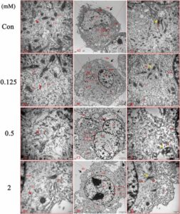
Our results showed that fluoride reduced the proliferation and survival rates of HT-22 cells. Cytomorphology showed that dendritic spines became shorter, cellular bodies became rounder, and adhesion decreased gradually after fluoride exposure. LDH results showed that fluoride exposure increased the membrane permeability of HT-22 cells. Transmission electron microscopy results showed that fluoride caused cells to swell, microvilli content decreased, cellular membrane integrity was damaged, chromatin was sparse, mitochondria ridge gap became wide, and microfilament and microtubule density decreased. Western Blot and qRT-PCR analyses showed that RhoA/ROCK/LIMK/Cofilin signaling pathway was activated by fluoride. F-actin/G-actin fluorescence intensity ratio remarkably increased in 0.125 and 0.5 mM NaF, and the mRNA expression of MAP2 was significantly decreased. Further studies showed that GLUT3 significantly increased in all fluoride groups, while GLUT1 decreased (p < 0.05). ATP contents remarkably increased, and ATP enzyme activity substantially decreased after NaF treatment with the control.
Conclusion
Fluoride activates the RhoA/ROCK/LIMK/Cofilin signaling pathway, impairs the ultrastructure, and depresses the connection of synapses in HT-22 cells. Moreover, fluoride exposure affects the expression of glucose transporters (GLUT1 and 3) and ATP synthesis. Sum up fluoride exposure disrupts actin homeostasis, ultimately affecting structure, and function in HT-22 cells. These findings support our previous hypothesis and provide a new perspective on the neurotoxic mechanism of fluorosis.
*Original full-text article online at: https://www.sciencedirect.com/science/article/pii/S0147651323002221