As demonstrated by the studies below, skeletal fluorosis may produce adverse symptoms, including arthritic pains, clinical osteoarthritis, gastrointestinal disturbances, and bone fragility, before the classic bone change of fluorosis (i.e., osteosclerosis in the spine and pelvis) is detectable by x-ray. Relying on x-rays, therefore, to diagnosis skeletal fluorosis will invariably fail to protect those individuals who are suffering from the pre-skeletal phase of the disease. Although the development of DXA bone density tests has enabled doctors to use a more sensitive test for bone density changes in the spine, DXA will fail to diagnose pre-skeletal fluorosis where the patient has not yet experienced a significant bone density effect. Moreover, some individuals with clinical skeletal fluorosis will not develop an increase in bone density, let alone osteosclerosis, of the spine. Thus, relying on unusual increases in spinal bone density will under-detect the rate of skeletal fluoride poisoning in a population.
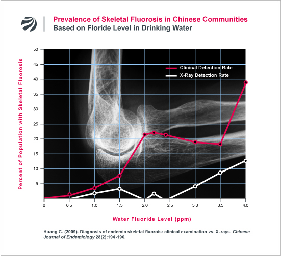
The problem with relying on x-ray changes for diagnosing skeletal fluorosis is highlighted by the data in the following figure. As can be seen, the clinical symptoms of skeletal fluorosis (e.g., restricted joint movement, joint pain, etc) occurred in many people that did not have fluorosis that was detectable by x-ray. (To read FAN’s translation of this study, click here).
pre-Skeletal Fluorosis:
“The radiological severity of knee osteoarthritis was greater in the endemic fluorosis group than in controls… [S]ome radiological findings such as osteosclerosis, interosseous membrane calcification, or ligament calcification, which are accepted as hallmarks of skeletal fluorosis were not found as frequently as in the literature.” (NOTE from FAN: In this group of fluorosis patients, only 3.6% had radiological evidence of osteosclerosis in the spine, and only 9% had evidence of calcification in the interosseous membrane of the forearm. Hence, the exacerbation of osteoarthritis occurred in most patients before the fluorosis was detectable on x-ray.)
SOURCE: Savas S, et al. (2001). Endemic fluorosis in Turkish patients: relationship with knee osteoarthritis. Rheumatology International 21: 30-5.
“Radiographs of the skeleton and bone scintigraphy showed degenerative osteoarthritis… Interestingly, laboratory findings, skeletal radiographs and bone densitometry, gave no indication for abnormalities of bone metabolism or mineralization. Without bone biopsy we would have failed the correct diagnosis (of skeletal fluorosis).”
SOURCE: Roschger P, et al. (1995). Bone mineral structure after six years fluoride treatment investigated by backscattered electron imaging (BSEI) and small angle x-ray scattering (SAXS): a case report. Bone 16:407.
“Assessment of the fluoride-induced changes from x-ray results is often difficult, especially in the initial stages commonly encountered.”
SOURCE: Czerwinski E, et al. (1988). Bone and joint pathology in fluoride-exposed workers. Archives of Environmental Health 43: 340-343.
“Ironically, two crucial criteria of fluorosis, i.e., osteosclerosis and bone pattern alteration, are the most questionable in the (x-ray) assessment. Perhaps this is one reason why such great discrepancies in the frequency of fluorosis are found among various authors.”
SOURCE: Czerwinski E, et al. (1988). Bone and joint pathology in fluoride-exposed workers. Archives of Environmental Health 43: 340-343.
“A wide variety of vague, subtle symptoms occurred either prior to or simultaneously with the development of bone changes similar to those reported previously. Nonskeletal symptoms, therefore, are important for early diagnosis.”
SOURCE: Zhiliang Y, et al. (1987). Industrial Fluoride Pollution in the Metallurgical Industry in China. Fluoride 20: 118-125.
“Arthritis of spine and small joints of hands and fingers develops early in the course of the disease with or without demonstrable radiological changes.”
SOURCE: Bhavsar BS, Desai VK, Mehta NR, Vashi RT, Krishnamachari KAVR. (1985). Neighborhood Fluorosis in Western India Part II: Population Study. Fluoride 18: 86-92.
“Our findings demonstrate a highly significant relationship between the frequency of back and neck surgery, fractures, symptoms of musculoskeletal disease and a past history of diseases of the bones and joints. In the absence of so-called classic fluorosis, a disease complex was established which involves much more than merely the radiologic appearance of dense bone. Since more stringent regulations in many countries have resulted in reduced exposure to fluorides, it is reasonable to examine workers and watch for these findings instead of waiting for dense bone to appear which is related to massive exposure to fluoride.”
SOURCE: Carnow BW, Conibear SA. (1981). Industrial fluorosis. Fluoride 14: 172-181.
“Similar findings of musculoskeletal changes without classical x-ray signs of fluorosis in workers exposed to high levels of fluorides have appeared in a number of other studies. Of special importance is the large prospective study by Zislin and Girskaya (1974). They followed 2738 workers from the time they first came to work in an aluminum smelter and compared them with 1700 others employed in a nonfluoride producing industry. They found that nonspecific bone changes, musculoskeletal symptoms and other findings antedate the classic x-ray changes of fluorosis in the bones by five to seven years and concluded that the changes of fluorosis described by Roholm represent the late stage of the disease.”
SOURCE: Carnow BW, Conibear SA. (1981). Industrial fluorosis. Fluoride 14: 172-181.
“In our opinion it is often difficult to appreciate the bone density because too many variables are involved such as radiograph penetration, influence of overlying soft tissues, etc.”
SOURCE: Boillat MA, et al. (1980). Radiological criteria of industrial fluorosis. Skeletal Radiology 5: 161-165.
“To our knowledge, [skeletal fluorosis] has not been described in the literature prior to the onset of the typical bone changes. This is not surprising since the intital stage, like that of many other kinds of chronic poisoning, develops slowly and insidiously with ill-defined complaints that are difficult to attribute to their cause. For instance, in lead poisoning the characteristic hallmarks are ‘lead line’ of gums and radial nerver paralysis; in chronic cadmium poisoning, one sees softening of bones. However, they are always precded or accompanied by a variety of subtle, inconspicuous symptoms of the kind encountered in incipient, chronic fluoride poisoning. Actually, subclinical poisoning can harm vast numbers of people before obvious clinical symptoms appear. These mulitple, hidden effects of slow poisoning pose a strong challenge to our current concepts of ‘safe limits’ of toxic substances in our environment.”
SOURCE: Waldbott GL, Lee JR. (1978). Toxicity from repeated low-grade exposure to hydrogen fluoride – Case report. Clinical Toxicology 13: 391-402.
“In addition to pain in the lower spine which is associated with radiological changes, patients with negative x-ray findings also complain of pain in the lumbar-sacral area, an indication that symptoms precede changes demonstrable by x-ray.”
SOURCE: Czerwinski E, Lankosz W. (1977). Fluoride-induced changes in 60 retired aluminum workers. Fluoride 10: 125-136.
“In early stages, fluorosis is usually associated only with stiffness, backache, and joint pains which may suggest the diagnosis of rheumatism, rheumatoid arthritis, ankylosing spondylitis and osteomalacia. At this stage the radiological findings of skeletal fluorosis may not be evident and therefore most of these cases are either misdiagnosed for other kinds of arthritis or the patients are treated symptomatically for pains of undetermined diagnosis (PUD). The majority of our patients had received treatment for rheumatoid arthritis and ankylosing spondylitis before they came under our observation.”
SOURCE: Teotia SPS, et al. (1976). Symposium on the Non-Skeletal Phase of Chronic Fluorosis: The Joints. Fluoride 9(1): 19-24.
“We also found patients with slight radiological changes (subtle signs or O-I) who complained of intense pains in the spine and in the large joints. On the other hand, some patients whose fluorosis was radiologically distinct were almost without complaints.”
SOURCE: Franke J, et al. (1975). Industrial fluorosis. Fluoride 8: 61-83.
“In several patients we failed to notice evidence of typical sclerosis in the radiogram. Instead, the picture of so-called ‘hypertrophic atrophy’ was found… It is likely that a previously existing osteoporosis is superimposed upon fluorosis or the predominance of the fluoride-induced bone resorption in conjunction with thickening of the statically loaded bone structure may be responsible.”
SOURCE: Franke J, et al. (1975). Industrial fluorosis. Fluoride 8: 61-83.
“Arthritis of the spinal column develops early in the disease with or without demonstrable radiological changes.”
SOURCE: Waldbott GL. (1974). The pre-skeletal phase of chronic fluorine intoxication. Fluoride 7:118-122.
“In spite of this distinctive clinical picture of advanced fluorosis, the earlier stages of the disease are more difficult to recognize. The initial symptoms are quite non-specific and not obviously linked to fluoride. The onset of fluorosis leads to tingling sensations in the hands and feet, pain similar to arthritic pain in the joints and the lower back, stiffness, and motor weakness. The first reliable diagnostic sign is increased bone density in X-ray examination, but in some early cases early bone changes are not radiologically detectable… The lack of a clear clinical picture of the early stages of fluorosis makes this disease easy to overlook or to misdiagnose, even in its relatively advanced stages.”
SOURCE: Groth, E. (1973), Two Issues of Science and Public Policy: Air Pollution Control in the San Francisco Bay Area, and Fluoridation of Community Water Supplies. Ph.D. Dissertation, Department of Biological Sciences, Stanford University, May 1973.
“It should also be noted that chronic fluorosis is not easily diagnosed, and that few physicians have ever seen a case. Three of the cases reported in the U.S. literature were not diagnosed until post-mortem examination revealed excessive fluoride content in the bone. It is possible that the disease may be occurring to some extent without having been recognized.”
SOURCE: Groth, E. (1973), Two Issues of Science and Public Policy: Air Pollution Control in the San Francisco Bay Area, and Fluoridation of Community Water Supplies. Ph.D. Dissertation, Department of Biological Sciences, Stanford University, May 1973.
“This case supports the premise that some forms of arthritis are related to sub-clinical fluorosis, i.e. fluorosis which is not sufficiently advanced to show the characteristic skeletal changes radiologically.”
SOURCE: Cook HA. (1972). Crippling fluorosis related to fluoride intake (case report). Fluoride 5: 209-213.
“Possibly some cases of pain diagnosed as rheumatism or arthritis may be due to subclinical fluorosis which is not radiologically demonstrable.”
SOURCE: Cook HA. (1971). Fluoride studies in a patient with arthritis. The Lancet 1: 817.
“There has been very little (research) done, especially in the realm of ‘borderline’ or subclinical toxicity. And yet, it is precisely in this area that knowledge of most importance to man could be brought to light. To this day, many investigators still think of fluorosis exclusively in terms of osteosclerosis, whether crippling or non-crippling. This attitude is no longer valid, because osteosclerosis is only one of many skeletal abnormalities that can be induced by fluoride.”
SOURCE: Marier JR, Rose D. (1971). Environmental fluoride. National Research Council of Canada, Publication No. 12,226, Ottawa.
“Whereas dental fluorosis is easily recognized, the skeletal involvement is not clinically obvious until the advanced stage of crippling fluorosis… Such early cases are usually in young adults whose only complaints are vague pains noted most frequently in the small joints of the hands and feet, in the knee joints and in the joints of the spine. These cases are frequent in the endemic area and may be misdiagnosed as rheumatoid or osteo arthritis.”
SOURCE: Singh A, Jolly SS. (1970). Fluorides and Human Health. World Health Organization. pp 239-240.
“The frequent lack of increased density or derangement of trabecular structure of bone in our cases and the nonspecificity of the alterations of thie spine make both of these changes bad criteria for the diagnosis of fluorosis. The more peripheral findings of exostosis, apposition of new bone, ossification of ligaments and tendon insertions and metastatic, aberrant growth of new bone seem much more specific and constant.”
SOURCE: Vischer TL, et al. (1970). Industrial fluorosis. In: TL Vischer, ed. (1970). Fluoride in Medicine. Hans Huber, Bern. pp. 96-105.
In the early stages of skeletal fluorosis, the “only complaints are vague pains noted most frequently in the small joints of hands and feet, the knee joints and those of the spine. Such cases are frequent in the endemic area and may be misdiagnosed as rheumatoid or osteoarthritis. Such symptoms may be present prior to the development of definite radiological signs. A study of the incidence of rheumatic disorders in areas where fluoridation has been in progress for a number of years would be of interest.”
SOURCE: Singh A, et al. (1963). Endemic fluorosis. Epidemiological, clinical and biochemical study of chronic fluoride intoxication in Punjab. Medicine 42: 229-246.
“It is apparent that small grossly recognizable deposits, areas of hyperplasia and perhaps beginning exostoses can be produced in the bones of small animals without their being detected by roentgenographic methods. That this may be equally true in the case of human beings cannot be claimed, but the point seems worthy of mention because in some instances X-ray photography is the only means used to detect evidence of industrial exposure to fluorides on the part of workmen. It seems probable that changes comparable to those seen in the animals may have escaped detection. It remains to be determined whether disability or limitation of movement in certain parts of the body may also occur before bone changes are demonstrable on the X-ray plate.”
SOURCE: Largent EJ, Machle W, Ferneau IF. (1943). Fluoride ingestion and bone changes in experimental animals. Journal of Industrial Hygiene and Toxicology 25: 396-408.
“incipient changes (1st phase) may be difficult to distinguish (via x-ray) from physiological variations.”
SOURCE: Roholm K. (1937). Fluoride intoxication: a clinical-hygienic study with a review of the literature and some experimental investigations. London: H.K. Lewis Ltd.

